Accepting new patients. For appointments call us at 617-732-5130
Request an AppointmentAccepting new patients. For appointments call us at 617-732-5130
Request an Appointment
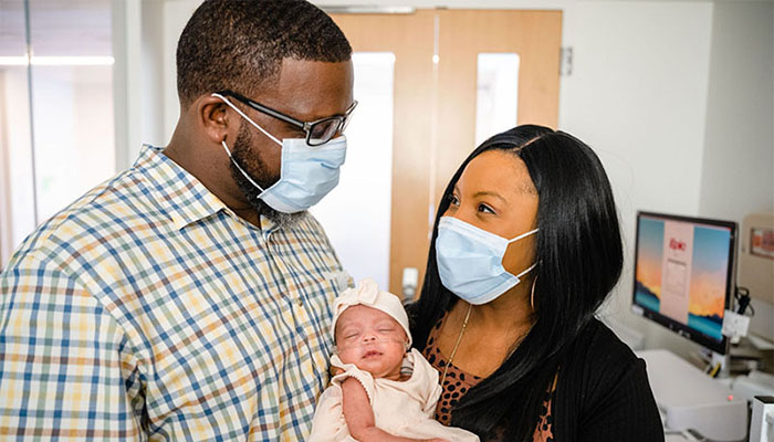
"I spend hours just looking at her in awe and saying, 'Wow, you really are a miracle,'" says Kenyatta Coleman, pictured in the Brigham Neonatal Intensive Care Unit (NICU) with husband Derek Coleman and their daughter, Denver, who became the first baby in North America to undergo in-utero brain surgery.
After having three children, Kenyatta Coleman was no stranger to unusual pregnancy symptoms. But when she began experiencing an intense itching on her leg while 28 weeks pregnant with her fourth child, she knew something wasn’t right.
She called her local obstetrician’s office in Baton Rouge, Louisiana for guidance. It was probably pregnancy hormones, they told her, suggesting that she try some over-the-counter antihistamines for relief.
But a mother’s intuition told Kenyatta that it was more serious, and she insisted on an appointment.
It was a decision that led her to the Maternal Fetal Medicine team at Brigham and Women’s Hospital and Boston Children’s Hospital in pursuit of care that would ultimately save her baby’s life.
During that local prenatal visit — which produced a diagnosis of cholestasis of pregnancy, the cause of her itchiness — her provider also noticed that Kenyatta was measuring smaller than expected, raising concerns about the baby’s growth and development.
An ultrasound at 30 weeks revealed an enlarged fetal heart, as well as a rare and deadly blood vessel abnormality inside her baby’s brain — a condition called vein of Galen malformation (VOGM) that occurs when misshapen arteries in the baby’s brain cause high-pressure blood to flow into the veins, leading to severe brain injury or loss of brain tissue.
When Kenyatta and her husband, Derek, learned that VOGM is typically only treated after the infant is born — when, in most cases, brain damage has already occurred, and the chance of survival is slim — they were heartbroken but determined to find another solution.
We didn’t know if we’d be bringing our baby home … She fought to be here.
Kenyatta and Derek traveled to Boston to participate in a highly innovative clinical trial to treat their baby’s condition with in-utero brain surgery. Performed by a multidisciplinary team of specialists from the Brigham in collaboration with Boston Children’s Hospital, it was the first procedure of its kind ever done in North America.
“We knew based on her scans that the size of the malformation, as well as how fast it was growing, meant that there was a very high chance of her going into immediate heart failure within the first two days of her life, so we were willing to do whatever was necessary,” Kenyatta said.
With seemingly impossible precision, the team repaired the misshapen blood vessels in the baby’s brain while she was still in the womb. The benefits were almost immediately apparent.
Baby Denver was born two days later, safe and healthy.
“We didn’t know if we’d be bringing our baby home. Now, here we are holding her in our arms and watching her meet her milestones,” Kenyatta said. “The outcome is mind-blowing. I spend hours just looking at her in awe and saying, ‘Wow, you really are a miracle.’ She fought to be here.”
The landmark fetal brain surgery was a collaborative effort by a team that included a maternal anesthesiologist, maternal-fetal medicine specialist, obstetrical radiologist, pediatric anesthesiologist, and pediatric neuro-interventional radiologist.
“In every fetal surgery, there are two patients — the baby and the mother — and caring for both is an important aspect of fetal procedures,” says Carol Benson, MD, chief of the Division of Ultrasound, co-director of High-Risk Obstetrical Ultrasound, and radiology director of the Vascular Laboratory at the Brigham. “You need to make sure that everything is aligned perfectly, and we couldn’t do anything without the precise communication and teamwork of everyone involved.”
Louise Wilkins-Haug, MD, PhD, division director of Maternal-Fetal Medicine and Reproductive Genetics at the Brigham, also emphasized the importance of this approach for such complex and delicate interventions.
“There may be three or four people at the table, but behind them there’s another dozen or so people assisting, and even more outside of the operating room who have been thinking about this from all angles,” Dr. Wilkins-Haug says.
The approach used to treat Denver in-utero involved placing tiny metal coils into the blood vessels in her brain through a catheter — inserted into the womb through a needle via ultrasound — to slow the blood flow and reduce pressure in the veins. Before the procedure began, a pediatric anesthesiologist administered medication for pain relief and to prevent her from moving during the procedure.

Baby Denver, pictured in the NICU with maternal-fetal medicine specialist Louise Wilkins-Haug, who helped perform the landmark procedure, received tiny metal coils in her brain while in the womb to correct a fatal abnormality.
After she was born, Denver stayed in the Brigham Neonatal Intensive Care Unit (NICU) for four weeks as the team closely monitored her progress. Soon enough, she was off medication, eating normally, and gaining weight.
Today, Denver continues to do well, and her brain shows no signs of any negative effects from VOGM, says Darren B. Orbach, MD, PhD, co-director of the Cerebrovascular Surgery and Interventions Center at Boston Children’s Hospital.
“We were thrilled that the aggressive decline usually seen after birth simply did not appear,” says Dr. Orbach, who inserted the coils through a catheter during the procedure. “While this is only our first treated patient, and it is vital that we continue the trial to assess the safety and efficacy in other patients, this approach has the potential to mark a paradigm shift in managing vein of Galen malformation — where we repair the malformation prior to birth and head off the heart failure before it occurs, rather than trying to reverse it after birth. This may markedly reduce the risk of long-term brain damage, disability, or death among these infants.”
While it is exciting to be part of a medical milestone, the greatest joy comes from seeing the impact on patients and families, said several members of the team.
“The malformation was growing so fast, but her brain was still OK, so this was a case where we could really help this little girl,” Dr. Benson says. “It’s very rewarding to know we are developing methods to help babies in ways that weren’t possible before.”
Having their unborn baby participate in an experimental surgery might give many parents-to-be pause, but the Colemans said they never once doubted they were making the best decision — one they say was guided equally by science and their strong religious faith as Baptists.
“We’re deeply rooted in our faith, and because of that we had people praying for us and we were also praying together,” Kenyatta says. “We told the team from the beginning, we’re here for a reason.”
As Kenyatta and Derek soak up this precious moment with the latest addition to their family, they also hope their story shines a light on the importance of third-trimester imaging to detect complications that develop late in pregnancy.
“I learned from my doctor later that I would have only had one ultrasound during my third trimester at 36 weeks,” Kenyatta said. “Based on the size of Denver’s malformation and what we knew about her heart function prior to the procedure, time was of the essence. Had we gone beyond 30 weeks, there is a possibility we might not have qualified for the intervention.”
Learn more about Maternal Fetal Medicine at Brigham and Women’s Hospital
For over a century, a leader in patient care, medical education and research, with expertise in virtually every specialty of medicine and surgery.
About BWH